Describe the mechanisms that confine cells and tissues to a specific anatomic site

Module 1 Assignment
Assignment Description:
Create a presentation addressing all of the following topics:
- Describe the mechanisms that confine cells and tissues to a specific anatomic site
- Discuss four types of cellular adaptations
- Compare and contrast necrosis and apoptosis
This PowerPoint® (Microsoft Office) or Impress® (Open Office) presentation should be a minimum of 15 slides (maximum of 20 slides), including a title, introduction, conclusion and reference slide, with detailed speaker notes and recorded audio comments for all content slides. Use the audio recording feature with the presentation software. Use at least four (4) scholarly sources and make certain to review the module’s rubric before starting your presentation.
Expert Answer and Explanation
Pathophysiology Cells and Cellular Adaptations
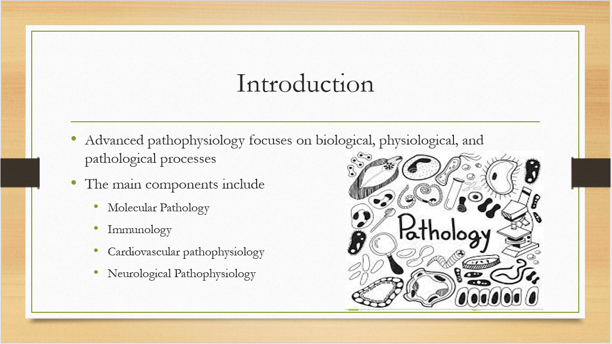
Advanced pathophysiology is a complex and in-depth field that studies the biological, physiological, and pathological processes underlying diseases and disorders.
Molecular pathology is a component of advanced pathophysiology that focuses on the molecular mechanisms underlying disease (Checa & Aran, 2020). This involves studying the genetic, biochemical, and cellular changes that occur in diseases at the molecular level. Immunology is the study of the body’s immune system and how it functions in response to disease.
Cardiovascular pathophysiology focuses on the biological processes that contribute to cardiovascular disease while Neurological pathophysiology addresses the structure and function of the brain and nervous system, as well as the mechanisms that contribute to disorders such as Alzheimer’s disease, Parkinson’s disease, and multiple sclerosis.
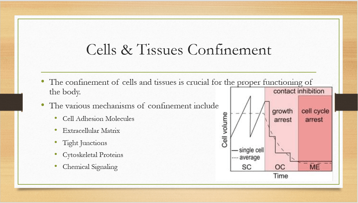
The confinement of cells and tissues to a specific anatomic site is crucial for the proper functioning of the body. There are several mechanisms that contribute to this confinement with each making use of different measures. Together, these mechanisms work in a coordinated manner to ensure the confinement of cells and tissues to specific anatomic sites, allowing the body to maintain its structural and functional integrity (Janiszewska et al., 2020).
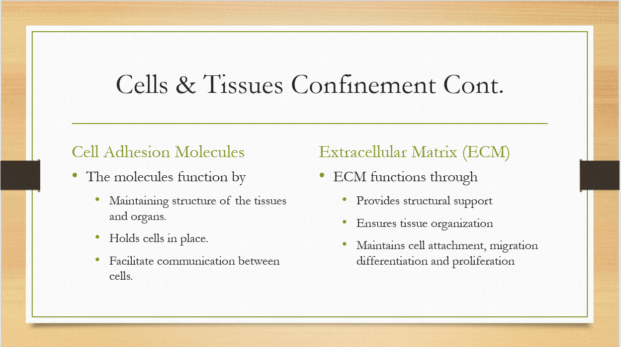
Cell adhesion molecules are proteins located on the cell surface that allow cells to bind to other cells or to the extracellular matrix (ECM). These molecules play a crucial role in maintaining the structure of tissues and organs by holding cells in place and allowing them to communicate with each other (Janiszewska et al., 2020).
On the other hand, The extracellular matrix (ECM) is a complex network of proteins and carbohydrates that surrounds cells and provides structural support. It plays a critical role in tissue organization and maintaining cell attachment, migration, proliferation, and differentiation (Janiszewska et al., 2020).
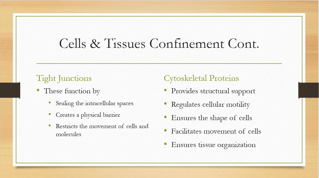
Tight junctions are specialized structures that seal the intercellular space between adjacent cells, creating a physical barrier that restricts the movement of molecules and cells between tissue compartments (Janiszewska et al., 2020). These junctions are particularly important in epithelial tissues, such as the skin and the lining of organs, where they prevent the diffusion of molecules and microbes into underlying tissues.
Additionally, the cytoskeleton is a complex network of proteins that provides structural support and regulates cellular motility. It includes three types of proteins: microfilaments, intermediate filaments, and microtubules. These proteins are responsible for the shape of cells, the organization of tissues, and the movement of cells (Janiszewska et al., 2020).
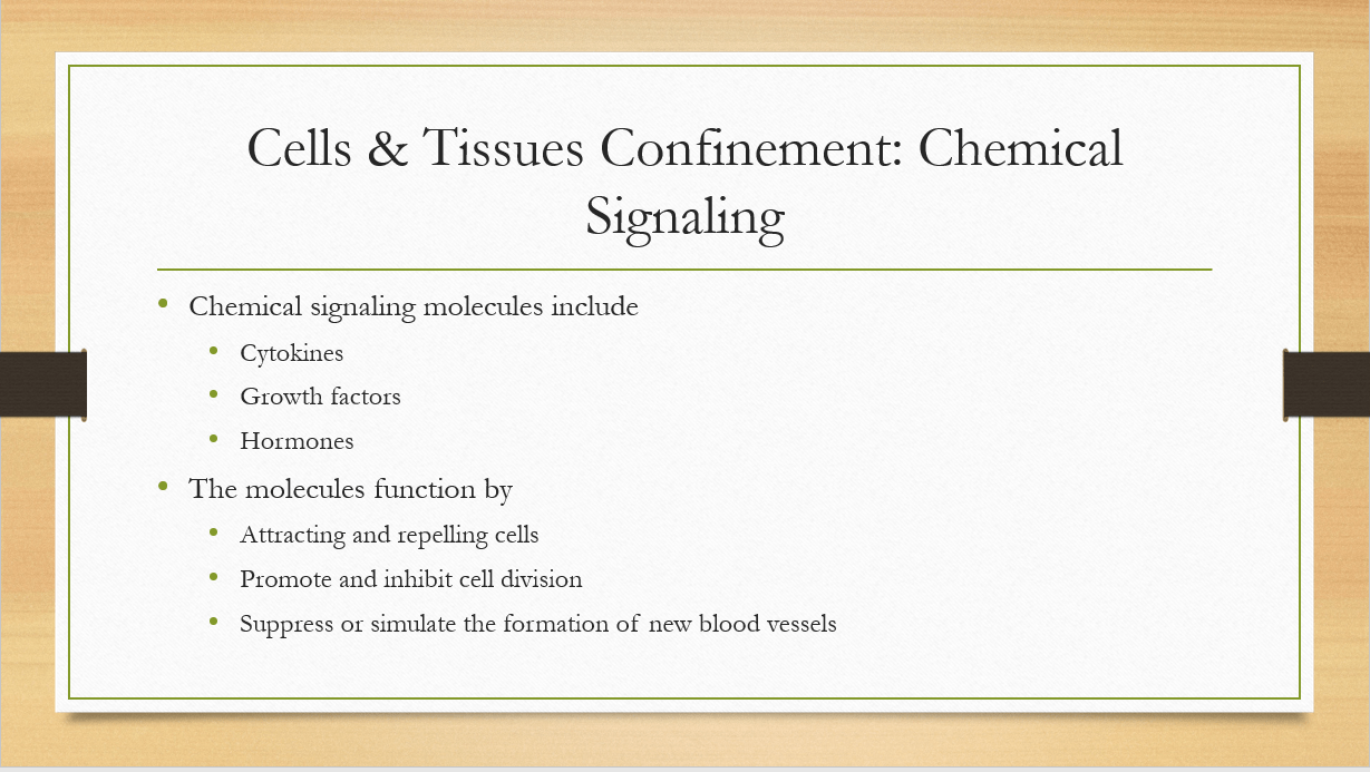
Chemical signaling molecules, such as growth factors, cytokines, and hormones, play a crucial role in regulating cell behavior and tissue organization. They can attract or repel cells, promote or inhibit cell division, and stimulate or suppress the formation of new blood vessels (Janiszewska et al., 2020).
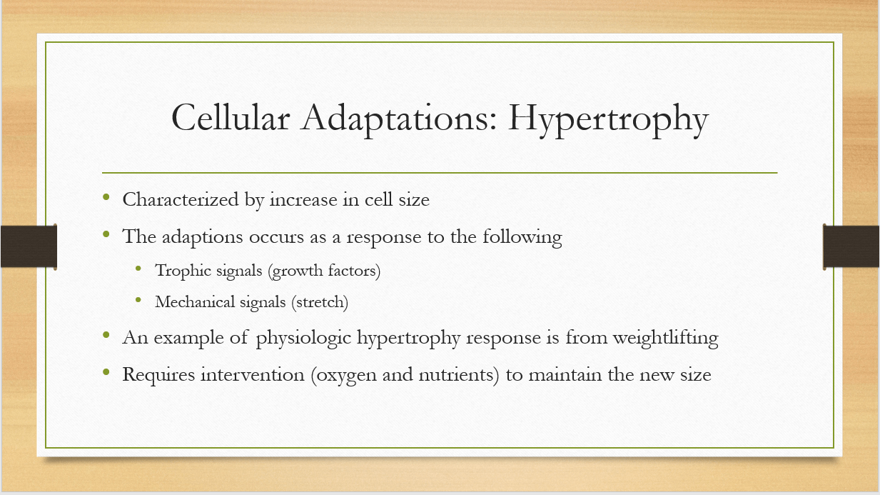
Hypertrophy is an increase in cell size, resulting in an increase in the size of the affected organ or tissue. This adaptation occurs in response to increased demand or stimulation, such as weightlifting causing an increase in muscle size or high blood pressure causing an increase in the size of the heart. The hypertrophied cells maintain their normal function and structure but require more nutrients and oxygen to support the increase in size (Lee et al., 2020).
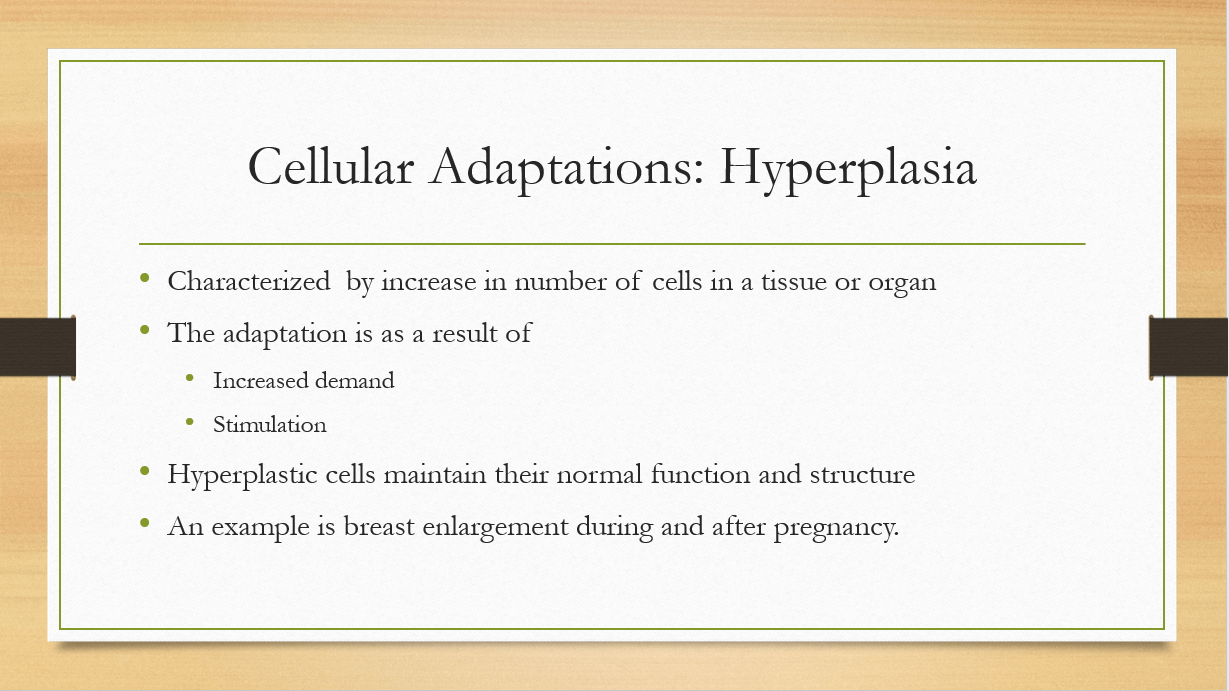
Hyperplasia is an increase in the number of cells in a tissue or organ, resulting in an increase in its size. This adaptation occurs in response to increased demand or stimulation, such as the proliferation of cells in the liver to compensate for the removal of part of the liver or the increase in breast tissue during pregnancy. The hyperplastic cells maintain their normal function and structure (Lee et al., 2020).
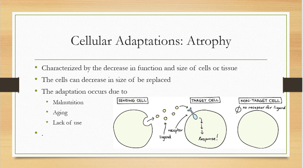
Atrophy is a decrease in the size and function of cells or tissue due to decreased demand or stimulation. This adaptation can occur due to a variety of factors, such as aging, lack of use, or malnutrition. The atrophied cells may decrease in size and function or be replaced by connective tissue (Lee et al., 2020).
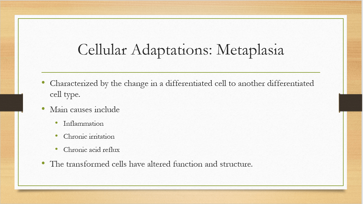
Metaplasia is a reversible change in the normal differentiated cell type of a tissue to another differentiated cell type. This adaptation occurs in response to chronic irritation or inflammation, such as chronic acid reflux causing the transformation of the normal esophageal lining to a type of epithelium more resistant to acid. The transformed cells may have altered structure and function and may be more susceptible to subsequent injury or disease (Lee et al., 2020).
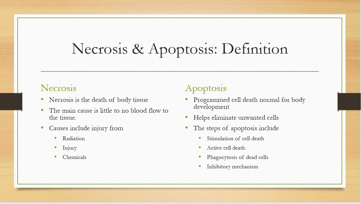
Necrosis is a type of cell death that occurs due to cellular injury, such as infection, toxins, or lack of oxygen. The main cause of necrosis is little to no blood flow to the tissue or organs as a result of injuries from injury, radiation, and chemicals (D’arcy, 2019).
Apoptosis is a type of programmed cell death that occurs as a normal part of the body’s development or to remove damaged or unnecessary cells. The main steps associated with apoptosis include stimulation of the cells, active cell death, phagocytosis of dead cells, and inhibitory mechanism (D’arcy, 2019).
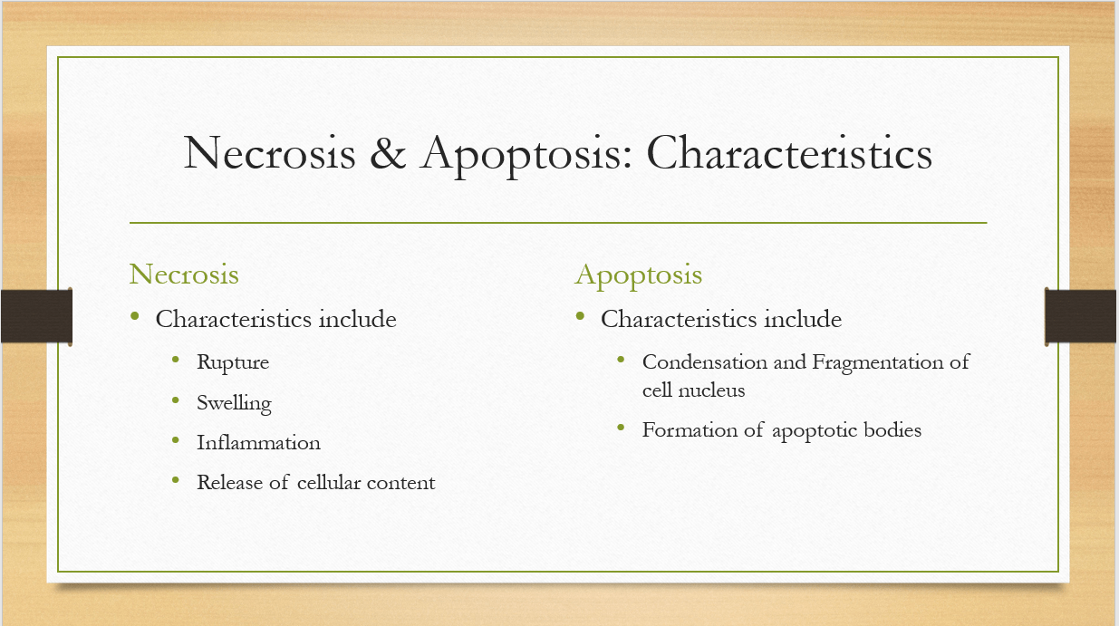
Necrosis is characterized by swelling, rupture, and release of cellular contents, which can cause inflammation and damage to nearby cells. Apoptosis is characterized by the condensation and fragmentation of the cell’s nucleus, and the formation of small membrane-bound vesicles called apoptotic bodies, which are then engulfed and digested by neighboring cells (D’arcy, 2019).
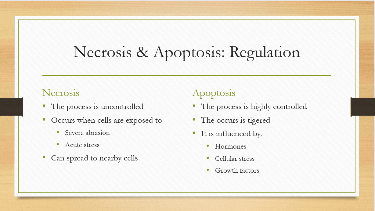
Necrosis is an uncontrolled process that occurs when cells are exposed to severe or acute stress, and can spread to nearby cells. Apoptosis is a highly regulated process that is triggered by specific signaling pathways and can be influenced by a variety of factors, such as hormones, growth factors, and cellular stress (D’arcy, 2019).
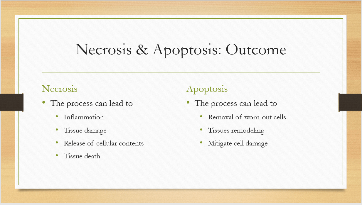
Necrosis can lead to tissue damage, inflammation, and the release of harmful cellular contents that can cause further injury and tissue death. Apoptosis results in the orderly removal of cells without causing tissue damage, and can be important in development, tissue remodeling, and the removal of damaged or potentially harmful cells (D’arcy, 2019).
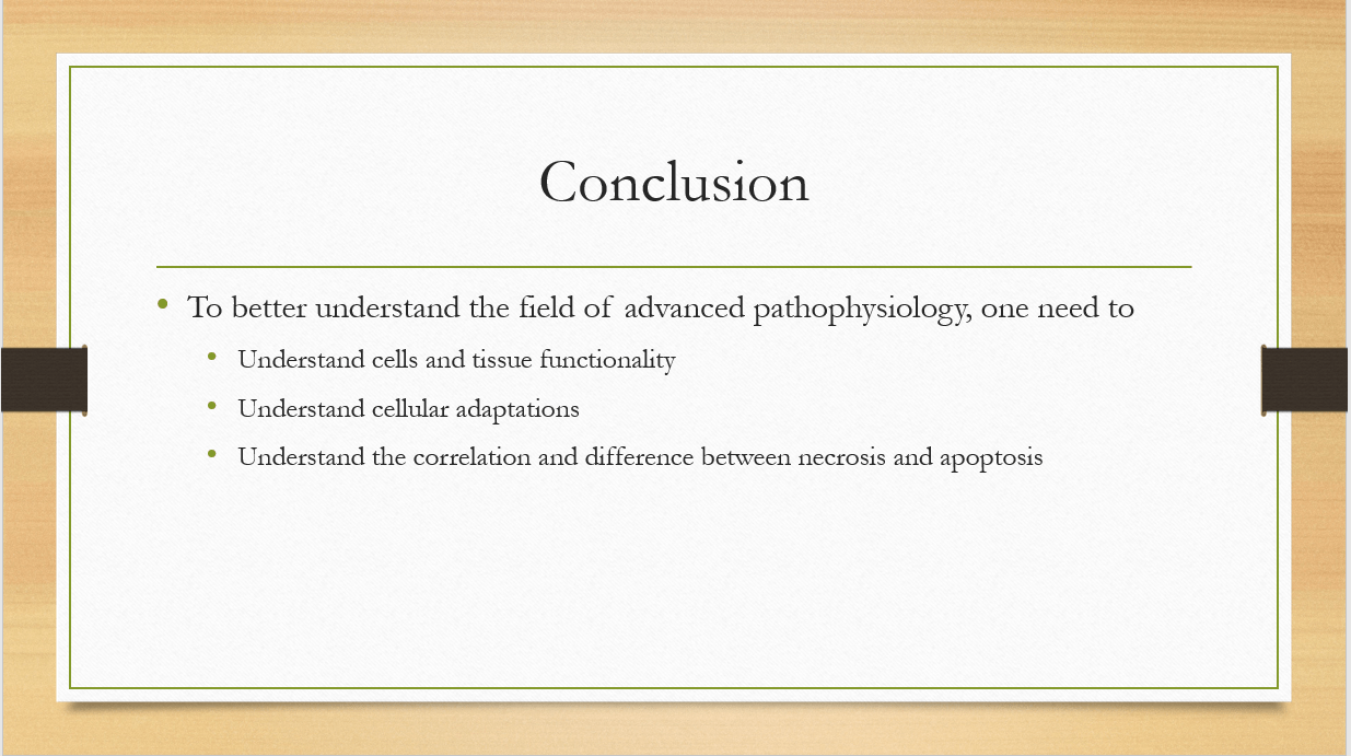
The different components and cell functionality are critical in the understanding of the field of study. The workings of the cell can be sued to help identify how the body responds to different stress or how they are structured. For instance, there are diseases that can affect tissues and organs and knowing the normal cell size if whether they are enlarged or diminished could tell if they are in the right state and functionality.
References
Checa, J., & Aran, J. M. (2020). Reactive oxygen species: drivers of physiological and pathological processes. Journal of Inflammation research, 1057-1073.
D’arcy, M. S. (2019). Cell death: a review of the major forms of apoptosis, necrosis and autophagy. Cell biology international, 43(6), 582-592.
Janiszewska, M., Primi, M. C., & Izard, T. (2020). Cell adhesion in cancer: Beyond the migration of single cells. Journal of Biological Chemistry, 295(8), 2495-2505.
Lee, P., Chandel, N. S., & Simon, M. C. (2020). Cellular adaptation to hypoxia through hypoxia inducible factors and beyond. Nature reviews Molecular cell biology, 21(5), 268-283.
Place your order now for a similar assignment and get fast, cheap and best quality work written by our expert level assignment writers. Use Coupon: NEW30 to Get 30% OFF Your First Order

Other Questions Related to this Class:
Module 1 Discussion
What is the relationship of environment and cancers? Describe the effect of tobacco, alcohol, radiation exposure, and diet and obesity to carcinogenesis.
Module 2 Discussion
Choose one disorder of the central or peripheral nervous system and discuss its clinical manifestations, prognosis, and pathophysiology.
Module 2 Assignment
Write a 1500-2000-word APA formatted essay of the following topics:
- Classify and differentiate at least four types of stroke
- Elaborate on the difference between primary and secondary Parkinson’s disease
- Summarize the five categories of pain. Discuss their pathways. Explain the etiology of chronic and acute pain
- Explain the pathophysiology and common clinical manifestations of Diabetes Mellitus Type II
Module 3 Discussion
Choose one of the following case studies from the Bruyere textbook and complete. Please post your answers, and then reply to two classmates.
Module 3 Assignment
Create a presentation addressing all of the following topics:
- Create a table differentiating the types of anemia, their clinical presentations, causes, and diagnostic tests
- What compensatory measures does the body employ in an attempt to restore cardiac output? What are the effect of these compensatory measures?
- Discuss the difference between right-sided and left-sided HF, their causes, clinical presentations, and diagnostic tests.
- How do the clinical presentations, prognosis, and management of acute and chronic leukemia differ?
This PowerPoint® (Microsoft Office) or Impress® (Open Office) presentation should be a minimum of 15 slides (maximum of 17 slides), including a title, introduction, conclusion and reference slide, with detailed speaker notes and recorded audio comments for all content slides. Use the audio recording feature with the presentation software. Use at least four scholarly sources and make certain to review the module’s Signature Assignment Rubric before starting your presentation.
Module 4 Discussion
Explain how cigarette smoking impacts the lungs at the physiologic level.
Module 4 Assignment
Write a 1500-2000 word APA formatted essay of the following topics:
- Discuss the pathophysiologic connection between asthma and allergies
- Discuss pathophysiology of lung cancer, clinical manifestations, and diagnostic tests
- What are the pathophysiologic changes in COPD and how does it differ from asthma?
- Discuss the use of oxygen therapy in patients with a diagnosis of COPD. What are the benefits and the potential pitfalls?
- Complete Case Study #13 (bacterial pneumonia) in the Bruyere textbook
Module 5 Discussion
Choose one of the following case studies from the Bruyere textbook and complete. Please post your answers, and then reply to two classmates.
Module 5 Assignment
Create a presentation addressing all of the following topics:
- Differentiate between osteoarthritis and rheumatoid arthritis. How does the pathophysiology of the diseases differ? How are they similar? What are the treatments for each disease?
- Explain the relationship between fluid and electrolyte balance and chronic kidney disease
- Explain the pathophysiology of the disease and discuss common clinical manifestations for chronic renal failure
- Differentiate between three types of muscle disorders
This PowerPoint® (Microsoft Office) or Impress® (Open Office) presentation should be a minimum of 17 slides (maximum of 20 slides), including a title, introduction, conclusion and reference slide, with detailed speaker notes and recorded audio comments for all content slides. Use the audio recording feature with the presentation software. Use at least four scholarly sources and make certain to review the module’s rubric before starting your presentation.
Module 6 Discussion
Choose one of the following case studies from the Bruyere textbook and complete. Please post your answers, and then reply to two classmates.
Module 6 Assignment
Write a 1500-2000 word APA formatted essay of the following topics:
- Explain the pathogenesis with common clinical presentation of celiacs disease
- Analyze the pathophysiology Crohn’s disease and relate genetic issues
- Differentiate between the five main infectious forms of hepatitis (A, B, C, D, E) from the two main non-infectious types of hepatitis (toxic and autoimmune)
- Elaborate on the pathogenesis and pathophysiology of pancreatic cancer
- Complete Case Study #26 (nausea and vomiting) in the Bruyere textbook
Module 7 Discussion
Choose one of the following biological agents and discuss epidemiology, pathogenesis, and safety issues. Then, in your response to a classmate discuss public health and vaccination considerations of your classmate’s chosen agent.
Module 7 Assignment
Write a 1500-2000 word APA formatted essay of the following topics:
- Differentiate among several types of shock: cardiogenic, hypovolemic, obstructive, distributive, and septic. Explain the pathophysiology of each.
- Discuss the risks of morbidity and mortality and the preparation and prevention measures for one type of natural disaster
- Explain how to set up for disaster closet in the emergency department. What items should it contain? Who should have access? Should it be locked? What type of drills should be conducted?
- Elaborate on the pathophysiology of burns. Differentiate between first, second, and third degree burns. What are the potential complications of each?
- Discuss the pathogenesis and progression of ebola. What safety concerns were addressed during the 2014 outbreak?
Module 8 Discussion
Choose one type of childhood cancer and discuss pathophysiology and prognosis of the disease.
Module 8 Assignment
Multiple Organ Failure (MOF) generally results from an infection, injury (surgical or accidental), or reduced blood flow which causes an uncontrolled inflammatory response. Identification and consideration to determine the underlying cause of MOF is essential. Though illness or injury may create multisystem issues and may impact many body systems, for this assignment focus on three body systems:
- Renal
- Pulmonary
- Circulatory
For this assignment, create a presentation for nursing staff education and discuss a specific illness, disorder or disease and the interrelationship with the renal, pulmonary, and circulatory systems, specifically identifying and describing how an alteration in one system may affect one or more of other two body systems. Furthermore, explain how the body tries maintain homeostasis and compensate for organ failure with assistance or reliance on another body system.
- Describe the illness, disorder or disease selected
- Characterize the interrelationship with the renal, pulmonary, and circulatory systems
- Identify & clarify how an alternation in one system impact the other two systems
- Describe how the body compensates for organ failure with assistance or reliance on another body system
Review the Signature Assignment Rubric before starting this assignment to ensure you are meeting all the essential requirements.
FAQs
What mechanisms confine cells and tissues?
The elegant architecture of life relies on a delicate balance between freedom and confinement. While individual cells need the flexibility to move, divide, and interact with their surroundings, they also require defined boundaries to maintain their shape, function, and organization within tissues. This essential balance is achieved through a fascinating interplay of various mechanisms that confine cells and tissues.
1. Extracellular Matrix (ECM): A Scaffolding Network
Imagine a city with intricate streets and avenues; the ECM plays a similar role for cells and tissues. This complex network of non-cellular proteins, such as collagen, fibronectin, and elastin, provides a structural scaffold and a biochemical environment for cells to adhere to, migrate through, and communicate with each other.
2. Cell-Cell Junctions: Building Bridges and Walls
Cells aren’t simply bricks stacked upon each other. They actively interact and communicate through specialized structures called cell-cell junctions. These junctions serve various purposes, including:
- Adherens junctions: These zipper-like connections, mediated by proteins like cadherins, act like strong glue, holding cells tightly together and forming continuous sheets of tissue.
- Tight junctions: Imagine watertight seals between cells; tight junctions prevent the leakage of molecules across cell membranes, maintaining distinct compartments within tissues and regulating transport between them.
- Gap junctions: These specialized channels allow direct communication between neighboring cells through the exchange of ions and small molecules, enabling coordinated tissue responses.
By forming these diverse connections, cells not only confine each other within tissues but also establish a dynamic communication network for coordinated function.
3. Basement Membranes: Anchoring Tissues
Think of a basement membrane as the foundation of a house, anchoring the tissue to the underlying structures. This specialized layer of ECM proteins forms a thin sheet beneath epithelial cells and serves several key functions:
- Physical support: The basement membrane provides mechanical strength and adhesion, anchoring the epithelium to the underlying connective tissue.
- Selective barrier: This layer acts as a filter, controlling the movement of molecules between the epithelium and the underlying tissues.
- Signaling hub: The basement membrane contains growth factors and signaling molecules that guide cell development, differentiation, and function.
4. Internal Cytoskeleton: A Push and Pull System
While external structures like the ECM and cell junctions provide physical confinement, the cell itself also possesses an internal scaffolding system called the cytoskeleton. This network of protein filaments, including microtubules, actin filaments, and intermediate filaments, plays a crucial role in:
- Maintaining cell shape: The cytoskeleton provides a supportive framework that resists deformation, helping cells maintain their defined boundaries within the tissue.
- Controlled movement: The dynamic assembly and disassembly of these filaments allow for cell migration, shaping, and division, all within the constraints of the surrounding tissue architecture.
- Organelle positioning: The cytoskeleton acts as tracks for transporting organelles within the cell, ensuring their proper placement and function within the confined space.
The coordinated interplay of these mechanisms, both external and internal, ensures the delicate balance between cellular freedom and confinement within tissues. This intricate dance is essential for maintaining tissue integrity, regulating cell behavior, and ultimately allowing organisms to function as a whole.
What is the difference between a cell and a tissue?
Cells and tissues, although both fundamental building blocks of life, differ in several key aspects:
Size and Complexity:
- Cells: Microscopic, individual units. Each cell is a complete living entity with its own membrane, internal structures (organelles), and the ability to carry out basic life functions like respiration, reproduction, and nutrient uptake.
- Tissues: Macroscopic, composed of groups of similar cells specialized for a specific function. They lack many of the internal structures and functions found in individual cells.
Organization and Independence:
- Cells: Relatively independent, able to perform some basic functions on their own. They can also specialize to a certain degree, but typically not as much as tissues.
- Tissues: Highly organized, with cells arranged in specific patterns and working together to perform a defined function. They depend on the coordinated activity of all cells within the tissue to function effectively.
Examples:
- Cells: Muscle cells, nerve cells, blood cells, skin cells, etc.
- Tissues: Muscle tissue, nervous tissue, epithelial tissue, connective tissue, etc.
Here’s a table summarizing the key differences:
| Feature | Cell | Tissue |
|---|---|---|
| Size | Microscopic | Macroscopic |
| Composition | Single unit | Groups of similar cells |
| Complexity | More complex with various organelles | Less complex, specialized for specific functions |
| Organization | Relatively independent | Highly organized, reliant on other cells |
| Examples | Muscle cells, nerve cells, blood cells, etc. | Muscle tissue, nervous tissue, epithelial tissue, etc. |
Remember, tissues are formed by groups of cells that have become specialized for a particular function. These cells often lose some of their independence and rely on the coordinated activity of the whole tissue to function effectively. This hierarchical organization, with cells forming tissues and tissues forming organs, allows for the complex functions and structures found in living organisms.
How do cells form tissues?
The journey from individual cells to organized tissues is a fascinating process orchestrated by a symphony of biological signals and cellular interactions. Here’s a breakdown of the key steps involved:
1. Cell Differentiation:
The first act begins with cell differentiation, where cells with similar gene expression potentials are guided to specialize for specific functions. This specialization is driven by various factors, including:
- Genetic cues: Specific genes are activated or repressed in different cells, leading to the expression of unique proteins and structures.
- Signaling molecules: Chemical messengers like hormones and growth factors bind to receptors on cells, triggering changes in gene expression and directing their development.
- Cell-cell interactions: Direct contact between cells can send signals and influence each other’s differentiation pathways.
As cells differentiate, they develop specific features like different shapes, protein production patterns, and organelle distributions, preparing them for their roles within the emerging tissue.
2. Cell Sorting and Migration:
Once specialized, cells embark on a choreographed dance, sorting themselves and migrating to their designated positions within the tissue. This intricate movement is guided by:
- Cell adhesion molecules: Specific proteins on cell surfaces act like Velcro, allowing cells of similar types to recognize and stick together.
- Chemotaxis: Certain chemicals act as signposts, attracting cells towards their proper positions within the tissue.
- Basement membranes: These specialized layers serve as tracks for cell migration and provide boundaries for tissue organization.
Through this dynamic sorting and migration, cells arrange themselves into specific patterns, paving the way for tissue formation.
3. Cell-Cell Junction Formation:
The final act involves building bridges and sealing walls. Cells establish crucial connections through various types of cell-cell junctions:
- Adherens junctions: These protein bridges, like sturdy zippers, bind cells tightly together, creating strong tissue cohesion.
- Tight junctions: Imagine watertight seals; these specialized junctions prevent leakage of molecules between cells, maintaining distinct compartments within the tissue.
- Gap junctions: These channels allow direct communication between neighboring cells through the exchange of ions and small molecules, enabling coordinated tissue responses.
By forming these diverse connections, cells not only secure their positions within the tissue but also establish a dynamic communication network for seamless collaboration.
4. Extracellular Matrix (ECM) Production:
As the tissue takes shape, cells begin weaving their own scaffolding – the extracellular matrix (ECM). This intricate network of non-cellular proteins like collagen, fibronectin, and elastin provides several critical functions:
- Structural support: The ECM acts as a supportive framework, giving the tissue its shape and mechanical strength.
- Biochemical environment: The ECM contains essential signaling molecules and growth factors that guide cell behavior and regulate tissue development.
- Selective barrier: The ECM controls the movement of molecules between cells and surrounding tissues.
The continuous production and remodeling of the ECM by cells further refines the tissue architecture and ensures its proper functioning.
This coordinated interplay of cell differentiation, sorting, junction formation, and ECM production ultimately leads to the formation of diverse and functional tissues. It’s a remarkable feat of cellular collaboration, essential for building the complex tapestry of life.
What name is given to a group of cells with similar structure and function?
There are two main terms used to describe a group of cells with similar structure and function:
-
Tissue: This is the most common and widely used term. A tissue is a collection of cells that have undergone the process of differentiation to become specialized for a specific function. Each type of tissue has its own characteristic structure and function. For example, muscle tissue is made up of elongated cells that can contract and relax, allowing for movement. Similarly, nervous tissue is composed of specialized cells called neurons that can transmit electrical signals, enabling communication throughout the body.
-
Epithelium: This term is often used specifically for tissues that cover the surface of the body or line its internal cavities and organs. Epithelial cells are tightly packed together and form a protective barrier against pathogens and harmful substances. Examples of epithelial tissues include skin, the lining of the digestive tract, and the inner lining of blood vessels.
While “tissue” is the broader term encompassing all groups of similar cells, “epithelium” refers to a specific type of tissue with a particular function and location.
So, depending on the context and the specific group of cells you’re referring to, either “tissue” or “epithelium” could be the appropriate term.

I am a professional nursing assignment expert offering comprehensive academic support to university nursing students across various institutions. My services are designed to help learners manage their workload effectively while maintaining academic excellence. With years of experience in nursing research, case study writing, and evidence-based reporting, I ensure every paper is original, well-researched, and aligned with current academic standards.
My goal is to provide dependable academic assistance that enables students to focus on practical training and career growth.
Contact me today to receive expert guidance and timely, high-quality nursing assignment help tailored to your academic needs.